
More Information
Submitted: October 30, 2026 | Accepted: November 04, 2026 | Published: November 06, 2026
Citation: Pu X, Wei L. A Behçet’s Disease Patient with Postoperative Perivalvular Leakage: A Case Report. J Clin Intensive Care Med. 2026; 11(1): 25-28. Available from:
https://dx.doi.org/10.29328/journal.jcicm.1001059
DOI: 10.29328/journal.jcicm.1001059
Copyright license: © 2026 Pu X, et al. This is an open access article distributed under the Creative Commons Attribution License, which permits unrestricted use, distribution, and reproduction in any medium, provided the original work is properly cited.
Keywords: Behçet’s disease; Echocardiography; Postoperative complications; Aortic valve; Paravalvular leak
Abbreviations: BD: Behçet’s Disease; CBD: Cardiovascular Behçet’s Disease; CT: Computed Tomography; CTA: Computed Tomography Angiography; ICBD: International Criteria for Behçet’s Disease; ISG: International Study Group; MRA: Magnetic Resonance Angiography; TAK: Takayasu Arteritis; AVR: Aortic Valve Replacement

A Behçet’s Disease Patient with Postoperative Perivalvular Leakage: A Case Report
Xinyu Pu1,3 and Liya Wei1*
1Department of Ultrasound, Peking University Shenzhen Hospital, Shenzhen, Guangdong, China
2Shantou University Medical College, Shantou, China
*Corresponding author: Liya Wei, Department of Ultrasound, Peking University Shenzhen Hospital, Shenzhen, Guangdong, China, Email: [email protected]
Behcet’s disease, a rare chronic recurrent vasculitis, affects various arteries and veins. A young female with aortic valve - involved Behcet’s disease had aortic valve replacement. Paravalvular leakage then occurred and quickly progressed to left ventricular wall dissection, leading to aortic valve root replacement. Current routine follow - up shows no significant issues. This article aims to boost clinicians’ awareness of Behcet’s disease for timely diagnosis and treatment of more patients.
Behçet’s disease (BD), a rare chronic relapsing vasculitis disorder, can affect various arteries and veins. Its main clinical features are oral or genital ulcers, uveitis, and skin lesions[1]. When involving the heart, it’s called cardiovascular Behcet’s disease (CBD). This article reports a CBD patient’s diagnosis and treatment to help clinicians better manage such cases.
A 24-year-old female developed chest tightness and dyspnea at 30 weeks of gestation, worsening on exertion, especially stair climbing. The patient has no prior history of cardiovascular diseases such as hypertension or coronary heart disease. She had a cesarean section at an external hospital at 31 weeks, but symptoms persisted postoperatively. She was transferred to our Cardiothoracic Surgery Department on April 8, 2017. Echocardiography showed aortic valve prolapse with severe regurgitation, periaortic abnormal echogenicity, and suspected perivalvular abscess (Figure 1). Antihypertensive therapy failed to halt disease progression. On April 21, 2017, emergency surgery was performed: “aortic sinus aneurysm repair (double-layer autologous pericardial patch), mitral valve repair, aortic valve replacement, tricuspid valve repair (DEVAGE technique)”. Intraoperatively, thickened aortic wall and rough endocardium suggested vasculitis. Postoperative pathology showed: Aortic valve fibrous thickening with fibrinoid necrosis, focal mucinous degeneration, and scattered lymphocytes; Aortic wall with fibrous capsule, adipose tissue, and lymphocytic infiltration. These features indicate sterile, immune-mediated inflammation rather than infection. Fibrinoid necrosis with mononuclear lymphocytic infiltration, without neutrophilic infiltration or identifiable microorganisms, supports vasculitis, and the lymphocytic infiltration of the aortic wall adipose tissue indicates active aortic wall inflammation consistent with BD vascular involvement. The postoperative patient’s symptoms of chest tightness and shortness of breath were relieved. Long-term anticoagulation treatment with “warfarin (maintained at 6 mg/day)” was continued, and “thalidomide 25 mg every night” was added.
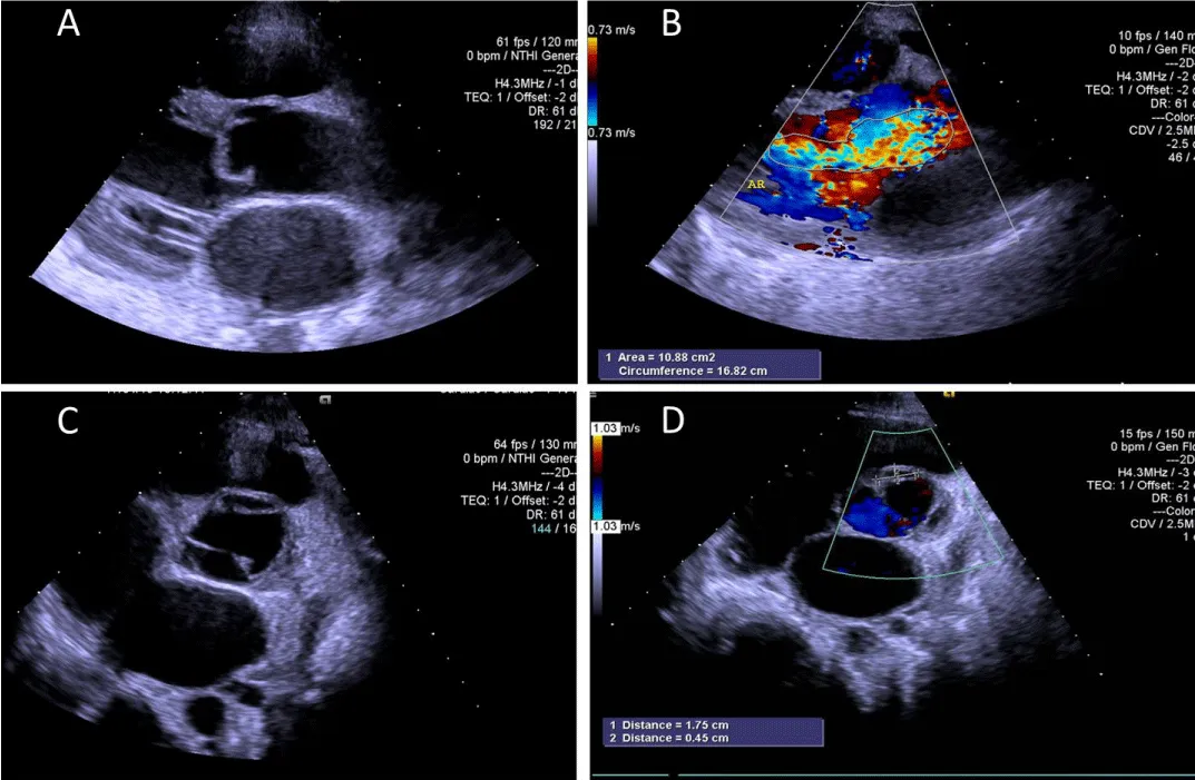
Figure 1: (A) Aortic valve prolapse with severe regurgitation. (B) Severe aortic valve regurgitation. (C, D) Periaortic valve abscess.suppression. Modified from Wilson et al., Nature Immunology. 2007;8(9):950–957.
On further medical history inquiry after surgery, the patient had recurrent oral ulcers starting in 2007, averaging 2 episodes per year, 2- 4 ulcers each time, mainly on the oral vestibule mucosa and tongue surface. The ulcers were large and painful, healing spontaneously within 2 weeks. There were also acne - like rashes, and no treatment was received. In 2016, the frequency of oral ulcers increased to 7- 8 times per year. Additionally, she had repeated vulvar ulcers, 7- 8 times per year, mostly on the labia minora, 2- 3 ulcers each time, with severe pain and spontaneous healing. During this period, she had intermittent right knee joint pain of unknown cause. The pain was more prominent after activity, relieved by rest, and resolved spontaneously within 1 week.
Based on the 2014 International Criteria for Behçet’s Disease (ICBD) (Table 1), considering the patient’s medical history, clinical manifestations, cardiac echocardiography, and intraoperative vascular conditions, the patient fulfilled the following items: recurrent oral aphthous ulcers (2 points), genital aphthous ulcers (2 points), acneiform skin lesions (1 point), and active aortic inflammation documented intraoperatively (1 point), giving a total ICBD score of 6 points, which exceeds the diagnostic threshold of ≥4 points. Accordingly, a final diagnosis of BD was made.
| Table 1: | |
| Symptoms | Score |
| Ocular lesions | 2 |
| Oral aphthosis | 2 |
| Genital aphthosis | 2 |
| Skin lesions | 1 |
| Central nervous system involvement | 1 |
| Vascular manifestations | 1 |
| Pathergy test (when used) | 1 |
| A patient scoring ≥4 points is classified as having BD | |
After a clear diagnosis of Behçet’s disease, according to the advice of the rheumatology and immunology doctor, “cyclophosphamide” was used for half a year (total dosage approximately 10 g), and then switched to “azathioprine (100 mg/day)”. The symptoms were well controlled. During the first year and a half after the surgery, regular cardiac color Doppler ultrasounds were conducted multiple times, and no obvious abnormalities were found.
On October 10, 2018, follow-up three-dimensional echocardiography revealed perivalvular leakage of the aortic mechanical valve (Figure 2A), and transesophageal echocardiography was performed for further evaluation (Figure 2B). Subsequent serial echocardiography consistently demonstrated moderate mechanical valve regurgitation (Figure 2C). On July 1, 2019, follow-up echocardiography identified left ventricular wall dissection communicating with the aorta through the gap between the valve frame and the anterior aortic wall, and surgical intervention was recommended (Figure 2D). Three days later, on July 4, 2019, computed tomography (CT) confirmed perivalvular leakage of the aortic mechanical valve and suggested possible left ventricular wall dissection (Figure 3). On July 19, 2019, CT at another hospital showed post-aortic valve replacement findings: a high-density annular shadow around the aortic valve with a fistula of approximately 9.2 mm; a hematoma-like protrusion measuring approximately 25.9 × 44.5 mm in the left anterior region of the aortic valve, compressing and narrowing the left ventricular outflow tract. Concurrent echocardiography showed severe perivalvular leakage of the mechanical valve and separation of the basal interventricular septum, consistent with interventricular septal dissection.
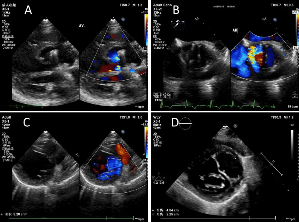
Figure 2: (A) Paravalvular leak of the aortic mechanical valve; (B) Paravalvular leak of the aortic mechanical valve detected by transesophageal echocardiography; (C) Moderate regurgitation in mechanical valve; (D) Formation of left ventricular wall dissection.
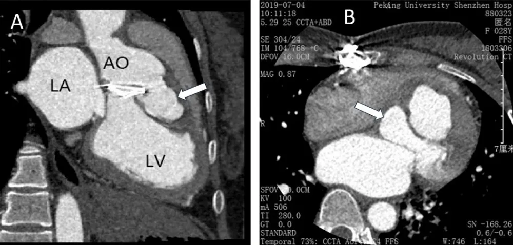
Figure 3: (A) Sagittal position; (B) Coronal position
On August 5, 2019, the patient underwent surgery at another hospital, including “ventricular septal aneurysm resection, aortic root replacement (Bentall procedure), and aortic valve paravalvular leakage repair”. Intraoperatively, coronary arteries were isolated and the existing aortic prosthetic valve removed. A 23# Regent valve was sutured to a 32# VESCUTEK prosthetic vessel to make a valved conduit, which was tailored into a septal patch for left ventricular outflow tract and aortic annulus reconstruction. The prosthetic valve was fixed to the annulus with interrupted sutures; the spacer was placed outside the aorta, with satisfactory knotting. Leaflets were continuously sutured to the residual aortic wall. Coronary arteries were circumferentially anastomosed to the prosthetic valve (5-0 polypropylene sutures), and the vessel’s distal end was end-to-end anastomosed to the aortic arch proximal end. Perioperatively, “methylprednisolone (1000 mg/day)” was given for 3 consecutive days. After discharge, the patient was followed up long-term in our outpatient clinic, with echocardiography every six months. Serial echocardiographic examinations showed no abnormalities through 2024.
BD is a systemic vasculitis first reported by Turkish physician Hulusi Behçet in 1937. It is also called “Silk Road disease” due to its high prevalence overlapping the ancient Silk Road [2]. Its etiology remains unclear, but it is thought to result from the interaction of internal and external factors including genetics, environment, innate and adaptive immunity [3].
BD presents with diverse clinical symptoms and lacks specific serological tests[4], making early diagnosis difficult. Clinically, the 2014 International Criteria for Behcet’s Disease (ICBD) (Table 1) and 1990 International Study Group (ISG) (Table 2) criteria are commonly used. While ICBD has slightly lower specificity (90.5%) than ISG (96.0%), its sensitivity (94.8%) is significantly higher than ISG (85.0%), highlighting its value in early identification [5]. Clinicians should detail symptoms like oral ulcers, genital ulcers, and skin lesions, and screen for immune-related diseases when encountering young and middle-aged patients with unexplained severe aortic regurgitation.
| Table 2: | |
| Clinical Manifestations | Definition |
| Recurrent oral ulcers | At least 3 recurrent episodes within 1 year |
| Plus any 2 of the following | |
| Recurrent genital ulcers | Genital aphthous ulcers or scars |
| Ocular lesions | Anterior and/or posterior uveitis, vitreous cells detected by slit-lamp examination, or retinal vasculitis observed by an ophthalmologist |
| Skin lesions | Erythema nodosum, pseudofolliculitis, papulopustular rash, or acneiform nodules in non-adolescent patients not taking corticosteroids |
| Positive pathergy test | Result evaluated by a physician 24–48 hours after the test |
TAK is a key differential for young females with aortic valve lesions. It is a chronic granulomatous vasculitis predominantly affecting women under 40, often involving the aortic valve to cause severe regurgitation and non-specific cardiac symptoms [6]. BD and TAK show differences in vascular phenotypes. TAK is mainly characterized by stenosis and occlusion, while BD is more commonly associated with aneurysms. In this case, the patient presented with an aortic sinus aneurysm rather than stenotic lesions, which is more consistent with the vascular phenotype of BD. Although both BD and TAK are vascular inflammatory lesions, TAK has rich imaging tools for early diagnosis: color Doppler ultrasound detects early aortic stenosis, diffuse wall thickening, or edema; contrast-enhanced ultrasound assesses wall neovascularization; CTA and MRA visualize large-vessel inflammation [7]. Therefore, the prognosis of TAK patients is significantly better than that of BD patients, and the risk of arterial dissection and rupture is higher in BD [8], possibly due to delayed diagnosis and insufficient inflammation control.
The aortic valve is the most commonly affected cardiac valve in BD, with regurgitation being the main manifestation. BD’s pathological hallmark is vasculitis [9], consistent with this patient’s postoperative pathology. Active aortic valve inflammation increases tissue fragility, thereby increasing surgical difficulty and the risk of paravalvular leakage. This patient had a complex history and critical condition, requiring urgent surgery without prior inflammation control. Intraoperatively, given the brittle aortic sinus and wall, weak annulus, thin leaflets, and poor myocardial function, a Bentall procedure was therefore not performed, and the aortic sinus aneurysm was repaired with a double-layer autologous pericardial patch combined with aortic valve replacement, mitral and tricuspid valve repair, and temporary pacing lead insertion. BD was diagnosed postoperatively, and the patient was started on regular immunosuppressive therapy and follow-up. Paravalvular leakage developed 1.5 years after surgery and progressed rapidly to left ventricular wall dissection at 2 years, which was later confirmed as interventricular septal dissection.
Previous studies have shown that the perioperative use of glucocorticoids, immunosuppressants, and biologics can control systemic inflammation and reduce vascular wall inflammation, thereby providing a more stable basis for surgery and potentially reducing the risk of complications such as perivalvular leakage and prosthetic valve detachment [10]. For surgical selection, the Bentall procedure has unique advantages over simple AVR. Resecting the diseased aortic root and annulus simultaneously allows direct healing between the outflow tract myocardium and mechanical valve ring. Additionally, its valve-carrying conduit exerts far less tension on the native annulus during movement than AVR’s mechanical valve, reducing traction on fragile tissue and promoting stable suture ring healing [11]. In this case, the two surgeries undergone by the patient are consistent with the aforementioned viewpoint. During the first surgery, due to changes in the patient’s condition, the operation was performed urgently while the inflammation had not been effectively controlled. Intraoperatively, the friable aortic tissue and weak annulus made a Bentall procedure inadvisable, and a root-preserving repair with aortic valve replacement was performed instead, resulting in paravalvular leak and interventricular septal dissection only 1.5 years postoperatively. In contrast, the second surgery was conducted with perioperative pulse methylprednisolone therapy to control vascular inflammation, which may have improved tissue conditions and facilitated completion of the Bentall procedure. Postoperatively, the patient remained stable and uneventful up to 2024.
Limitations of the case report
This report has several limitations that should be acknowledged. First, it describes the clinical course of a single patient; therefore, the findings cannot be generalized to all patients with cardiovascular Behçet’s disease. Second, although the clinical manifestations, imaging findings, and histopathological observations supported the diagnosis of Behçet’s disease, the available information regarding the microbiological investigations used to exclude infective endocarditis is limited. Third, some details of the patient’s treatment course, including the precise duration of immunosuppressive therapy and the relationship between inflammatory disease activity and the development of postoperative complications, could not be fully established from the available clinical records. Furthermore, the second surgical intervention was performed at another hospital, which may have limited access to certain detailed operative and postoperative records. Although the patient remained clinically stable during follow-up through 2024, longer-term observations would be valuable for assessing the durability of the surgical outcome and the risk of recurrent cardiovascular complications. Despite these limitations, this case provides useful clinical insights into the diagnostic challenges, surgical management, and postoperative complications associated with cardiovascular Behçet’s disease.
Cardiac Behçet’s disease (CBD) accounts for 1% - 6% of all BD cases but carries threefold higher mortality than non-cardiac BD [12]. Early diagnosis, perioperative management, and surgical strategy are pivotal for CBD prognosis, efficacy, and mortality reduction. Young-middle-aged patients with unexplained heart valve disease should prioritize ICBD screening to confirm/exclude CBD. For surgically indicated CBD, the Bentall procedure is first-line. Surgery should be deferred until BD is medically controlled to minimize risks and optimize recovery.
Acknowledgement
This research was supported by the Sanming Project of Medicine in Shenzhen (No. SZSM20211011), the Scientific Research Project of Peking University Shenzhen Hospital (No. LCYJ2021027), and the Shenzhen Science and Technology Program (No. JCYJ20210324105614038).
- Lavalle S, Caruso S, Foti R, Gagliano C, Cocuzza S, La Via L, et al. Behçet’s Disease, Pathogenesis, Clinical Features, and Treatment Approaches: A Comprehensive Review. Medicina (Kaunas) 2024;60:562. Available from: https://doi.org/10.3390/medicina60040562.
- Bergamo S, di Meo N, Stinco G, Bonin S, Trevisini S, Trevisan G. Adamantiades-Behçet Disease at the Beginning of the Silk Route: North-East Italian Experience. Acta Dermatovenerol Croat 2017;25:295–7.
- Yazici Y, Hatemi G, Bodaghi B, Cheon JH, Suzuki N, Ambrose N, et al. Behçet syndrome. Nat Rev Dis Primers 2021;7:67. Available from: https://doi.org/10.1038/s41572-021-00301-1.
- Hassan F, Jeries H, Naffaa ME. Challenges in the Timely Diagnosis of Behcet’s Disease. Life (Basel) 2023;13:1157. Available from: https://doi.org/10.3390/life13051157.
- International Team for the Revision of the International Criteria for Behçet’s Disease (ITR-ICBD). The International Criteria for Behçet’s Disease (ICBD): a collaborative study of 27 countries on the sensitivity and specificity of the new criteria. J Eur Acad Dermatol Venereol 2014;28:338–47. Available from: https://doi.org/10.1111/jdv.12107.
- Zhang Y, Yang K, Meng X, Tian T, Fan P, Zhang H, et al. Cardiac Valve Involvement in Takayasu Arteritis Is Common: A Retrospective Study of 1,069 Patients Over 25 Years. The American Journal of the Medical Sciences 2018;356:357–64. Available from: https://doi.org/10.1016/j.amjms.2018.06.021.
- Barra L, Kanji T, Malette J, Pagnoux C. Imaging modalities for the diagnosis and disease activity assessment of Takayasu’s arteritis: A systematic review and meta-analysis. Autoimmunity Reviews 2018;17:175–87. Available from: https://doi.org/10.1016/j.autrev.2017.11.021.
- Choi SJ, Koo HJ, Yang DH, Kang JW, Oh JS, Hong S, et al. Comparison of Clinical, Angiographic Features and Outcome in Takayasu’s Arteritis and Behçet’s Disease With Arterial Involvement. J Rheum Dis 2020;27:100. Available from: https://doi.org/10.4078/jrd.2020.27.2.100.
- Fei Y, Li X, Lin S, Song X, Wu Q, Zhu Y, et al. Major vascular involvement in Behçet’s disease: a retrospective study of 796 patients. Clin Rheumatol 2013;32:845–52. Available from: https://doi.org/10.1007/s10067-013-2205-7.
- Kageyama S, Ohashi T, Tadakoshi M, Kamikawa Y, Kojima A, Yamauchi H, et al. [Four Times Aortic Valve Replacement for Recurrent Aortic Annulus Destruction Due to Behçet’s Disease: Report of a Case]. Kyobu Geka 2021;74:681–5.
- Jung Y, Ahn BH, Lee KS, Jeong IS, Kim KH, Na KJ, et al. A novel solution to prosthetic valve dehiscence after aortic valve surgery in Behçet’s disease. Interact Cardiovasc Thorac Surg 2017;24:342–7. Available from: https://doi.org/10.1093/icvts/ivw361.
- Geri G, Wechsler B, Thi Huong DL, Isnard R, Piette J-C, Amoura Z, et al. Spectrum of Cardiac Lesions in Behçet Disease: A Series of 52 Patients and Review of the Literature. Medicine 2012;91:25–34. Available from: https://doi.org/10.1097/MD.0b013e3182428f49.